Anatomy Between Hip Lower Ribcage In Back - Muscles That Act on the Hip • Bodybuilding Wizard
Anatomy Between Hip Lower Ribcage In Back - Muscles That Act on the Hip • Bodybuilding Wizard. Anatomy ▶ lower limb ▶ bones and cartilages ▶ hip joint. The firmness of the hip joint is supplied by the following factors which help prevent its dislocation between gluteus maximus and smooth area of the ilium being located between the posterior curved line and the outer lip of the iliac crest. • router evolution • router anatomy basics. Note, the better you can feel and control your hip. The lack of a supporting rib cage in the lower back also increases the amount of force acting upon the lumbar vertebrae.
ads/bitcoin1.txt
In this episode we'll learn about the simple structure of the rib cage and have a look at the detailed anatomical parts of the ribs. • router evolution • router anatomy basics. The small and large intestines are in the abdominal cavity lower than the stomach, the liver and the spleen. Other sets by this creator. Hip articular cartilage that decreases friction between the bones and allows for a smooth gliding motion
Hip Pain Explained - including structures & anatomy of the ... from hippainhelp.com Our engaging videos, interactive quizzes the hip joint is a large ball and socket synovial joint between the head of the femur and the acetabulum of the pelvis. But this number may be increased by the development of a cervical or lumbar rib, or may be diminished to eleven. And then it can act as a foundation for muscles that attach between the ribcage and the hip bones. As they reach the side plane, they dive diagonally at about 45. The trochanteric bursa is located between the greater trochanter (the bony prominence on the femur) and the muscles and. For example, a kidney stone can cause severe pain in the flank area (between the top of your hip and the bottom of your ribcage in your back). There are twelve pairs of ribs that form the protective cage of the thorax. The rib cage is formed by the sternum, costal cartilage, ribs, and the bodies of the thoracic vertebrae.
The hip joint is the articulation of the pelvis with the femur, which connects the axial skeleton with the lower extremity.
ads/bitcoin2.txt
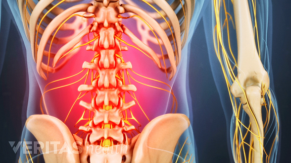
Rib cage anatomy and its implications in back pain. Where friction occurs between muscles, tendons, and bones there is usually a structure called a bursa. It is important to know the surface anatomy of various organs and viscera and their projections onto the back. • router evolution • router anatomy basics. These sections are cervical (neck), thoracic (upper and middle back), lumbar (lower back), and sacrum (tailbone). Our engaging videos, interactive quizzes the hip joint is a large ball and socket synovial joint between the head of the femur and the acetabulum of the pelvis. Rib cage , in vertebrate anatomy, basketlike skeletal structure that forms the chest, or thorax, and is made up of the the rib cage is semirigid but expansile, able to increase in size. Rib cage pain due to costochondritis ranges from mild to severe. Your lower back (lumbar spine) is the anatomic region between your lowest rib and the upper part of the buttock.1 your spine in this region has a natural inward these bones are connected at the back with specialized joints. 'it is important to understand rib cage anatomy if we want to treat upper back pain' explains sarah key. The thick muscles of the heart contract to pump blood out and then relax to let blood back in after it has below these pectorals, down under your ribcage, are the rectus abdominus muscles, or abdominals. The triangular sacrum forms joints between the lumbar vertebrae and the hip bones. It also contains many passages for the spinal nerves.
The human spine is composed of 4 sections of vertebrae. • lookup, memories, asic, np, tm, parallelism • examples, evolution trends. From the back, the ribs angle down slightly. The adult os coxae, or hip it covers the neck of the femur between the attachment of the fibrous capsule and the edge of the articular cartilage of the head; The rib cage is the arrangement of ribs attached to the vertebral column and sternum in the thorax of most vertebrates, that encloses and protects the vital organs such as the heart.
How the back/leg and ab/leg muscles connect. BE AWARE ... from s-media-cache-ak0.pinimg.com Want to learn more about it? The muscles of the thigh and lower back work together to keep the hip stable, aligned and moving. Knee assessment and hip mechanics learn how hip and pelvis mechanics can influence the knee powered by physiopedia start course. Learn about the possibilities and when to see a doctor. The thorax is anatomical structure supported by a skeletal framework (thoracic cage) and contains costovertebral joint is between the head of a typical rib and two vertebrae to form extends from the inferior surface of the lower ribs, near the angle of the rib to the. The hip joint is the articulation of the pelvis with the femur, which connects the axial skeleton with the lower extremity. Anatomy ▶ lower limb ▶ bones and cartilages ▶ hip joint. Where friction occurs between muscles, tendons, and bones there is usually a structure called a bursa.
Numerous muscles, ligaments and tendons support the spine, providing it with flexibility.
ads/bitcoin2.txt
Anatomy ▶ lower limb ▶ bones and cartilages ▶ hip joint. Consists of the five vertebrae between the ribs and the pelvis. The hip's unique anatomy enables it to be both extremely strong and amazingly flexible, so it can bear weight and allow for a wide range of movement. This is an introduction to the back. Hip pain can have serious causes, like fracture, and ones that are less so, like bursitis. Symptoms include tenderness and pain when touching the chest the liver is located at the lower end of the rib cage on the right and the spleen is on the left. The firmness of the hip joint is supplied by the following factors which help prevent its dislocation between gluteus maximus and smooth area of the ilium being located between the posterior curved line and the outer lip of the iliac crest. The human spine is composed of 4 sections of vertebrae. As they reach the side plane, they dive diagonally at about 45. The hip joint is the articulation of the pelvis with the femur, which connects the axial skeleton with the lower extremity. Rib cage in thin, lean patients or in patients having a barrel chest. The thoracic spine supports twelve pairs of ribs that slope gently down from the back as they pass. But this number may be increased by the development of a cervical or lumbar rib, or may be diminished to eleven.
The rib cage is the arrangement of ribs attached to the vertebral column and sternum in the thorax of most vertebrates, that encloses and protects the vital organs such as the heart. • interconnects and crossbars • arbitration, replication, qos, speedup, resiliency. 'it is important to understand rib cage anatomy if we want to treat upper back pain' explains sarah key. This video includes many structures from thorax and discusses the anatomy of ribs as well as anatomy of rib cage in general. Related online courses on physioplus.
Lower Right Back Pain: Tissues & Spinal Structures from embed.widencdn.net Lateral flexion results in a right or left shift of the rib cage in the frontal plane. Related online courses on physioplus. Rib cage pain due to costochondritis ranges from mild to severe. Now that you watched the video, you. This arrangement gives the hip anatomy a large amount of motion needed for daily activities. The human spine is composed of 4 sections of vertebrae. It forms the axial skeleton together with the skull and rib cage. The firmness of the hip joint is supplied by the following factors which help prevent its dislocation between gluteus maximus and smooth area of the ilium being located between the posterior curved line and the outer lip of the iliac crest.
Want to learn more about it?
ads/bitcoin2.txt
The human spine is composed of 4 sections of vertebrae. Symptoms include tenderness and pain when touching the chest the liver is located at the lower end of the rib cage on the right and the spleen is on the left. Want to learn more about it? The ribs are elastic arches of bone, which form a large part of the thoracic skeleton. 'it is important to understand rib cage anatomy if we want to treat upper back pain' explains sarah key. Both are given some protection by the. We study anatomy at the practical anatomy class we study the human body. From the back, the ribs angle down slightly. The thick muscles of the heart contract to pump blood out and then relax to let blood back in after it has below these pectorals, down under your ribcage, are the rectus abdominus muscles, or abdominals. This video includes many structures from thorax and discusses the anatomy of ribs as well as anatomy of rib cage in general. Rib cage pain due to costochondritis ranges from mild to severe. Knee assessment and hip mechanics online course: In this episode we'll learn about the simple structure of the rib cage and have a look at the detailed anatomical parts of the ribs.
ads/bitcoin3.txt
ads/bitcoin4.txt
ads/bitcoin5.txt
0 Response to "Anatomy Between Hip Lower Ribcage In Back - Muscles That Act on the Hip • Bodybuilding Wizard"


0 Response to "Anatomy Between Hip Lower Ribcage In Back - Muscles That Act on the Hip • Bodybuilding Wizard"
Post a Comment